UFGI publication round-up week 5/22/17
The nucleus is irreversibly shaped by motion of cell boundaries in cancer and non-cancer cells.
Author information: Tocco VJ1, Li Y1, Christopher KG1, Matthews JH2, Aggarwal V1, Paschall L1, Luesch H2, Licht JD3, Dickinson RB1, Lele TP1,4.
Date of e-pub: May 2017
Abstract: Actomyosin stress fibers impinge on the nucleus and can exert compressive forces on it. These compressive forces have been proposed to elongate nuclei in fibroblasts, and lead to abnormally shaped nuclei in cancer cells. In these models, the elongated or flattened nuclear shape is proposed to store elastic energy. However, we found that deformed shapes of nuclei are unchanged even after removal of the cell with micro-dissection, both for smooth, elongated nuclei in fibroblasts and abnormally shaped nuclei in breast cancer cells. The lack of shape relaxation implies that the nuclear shape in spread cells does not store any elastic energy, and the cellular stresses that deform the nucleus are dissipative, not static. During cell spreading, the deviation of the nucleus from a convex shape increased in MDA-MB-231 cancer cells, but decreased in MCF-10A cells. Tracking changes of nuclear and cellular shape on micropatterned substrata revealed that fibroblast nuclei deform only during deformations in cell shape and only in the direction of nearby moving cell boundaries. We propose that motion of cell boundaries exert a stress on the nucleus, which allows the nucleus to mimic cell shape. The lack of elastic energy in the nuclear shape suggests that nuclear shape changes in cells occur at constant surface area and volume. This article is protected by copyright. All rights reserved.
Metal transporter ZIP14 (SLC39A14) deletion in mice increases manganese deposition and produces neurotoxic signatures and diminished motor activity.
Author information: Aydemir TB1, Kim MH1, Kim J1, Colon-Perez LM2,3, Banan G3, Mareci TH3,4, Febo M2,3, Cousins RJ5,4.
Date of e-pub: May 2017
Abstract: Mutations in human ZIP14 have been linked to symptoms of early onset of Parkinsonism and Dystonia. This phenotype is likely related to excess manganese accumulation in the central nervous system. The metal transporter ZIP14 (SLC39A14) is viewed primarily as a zinc transporter that is inducible via proinflammatory stimuli. In vitro evidence shows that ZIP14 can also transport manganese. To examine role for ZIP14 in manganese homeostasis, we used Zip14 knockout (KO) male and female mice to conduct comparative metabolic, imaging and functional studies. Manganese accumulation was 4-5 fold higher in brain of Zip14 KO mice compared to young adult wild type (WT) mice. Accumulation of subcutaneously administered 54Mn was less in liver, gallbladder and gastrointestinal tract of the KO mice suggesting manganese elimination is impaired with Zip14 ablation. Impaired elimination creates the opportunity for atypical manganese accumulation in tissues including the brain. Intensity of MRI images from brains of the Zip14 KO mice are indicative of major manganese accumulation. Ingreement with excessive manganese accumulation was impaired motor function observed in the Zip14 KO mice. These results also demonstrate that ZIP14 is not essential for manganese uptake by the brain. Nevertheless, the upregulation of signatures of brain injury observed in the Zip14 KO mice demonstrates normal ZIP14 function is an essential factor required to prevent manganese-linked neurodegeneration.
Manganese is an essential micronutrient. When acquired in excess manganese accumulates in tissues of the central nervous system and is associated with neurodegenerative disease, particularly Parkinson-like syndrome and Dystonia. Some members of the ZIP metal transporter family transport manganese. Using mutant mice deficient in the ZIP14 metal transporter, we have discovered that ZIP14 is essential for manganese elimination via the gastrointestinal tract and a lack of ZIP14 results in manganese accumulation in critical tissues such as the brain as measured by MRI and produces signatures of brain injury and impaired motor function. Humans with altered ZIP14 function would lack this gatekeeper function of ZIP14 and therefore would be prone to manganese-related neurological diseases.
*Corrections have been made according to the original information from the authors.
A Flow Cytometry-Based Screen Identifies MBNL1 Modulators that Rescue Splicing Defects in Myotonic Dystrophy Type I.
Author information: Zhang F1, Bodycombe NE2, Haskell KM3, Sun YL1, Wang ET4, Morris CA1,5, Jones LH2,6, Wood LD1, Pletcher MT1,7.
Date of e-pub: May 2017
Abstract: Myotonic Dystrophy Type 1 (DM1) is a rare genetic disease caused by expansion of CTG trinucleotide repeats ((CTG)exp) in the 3′ untranslated region of the DMPK gene. The repeat transcripts sequester the RNA binding protein Muscleblind-like protein 1 (MBNL1) and hamper its normal function in pre-mRNA splicing. Overexpressing exogenous MBNL1 in the DM1 mouse model has been shown to rescue the splicing defects and reverse myotonia. Although a viable therapeutic strategy, pharmacological modulators of MBNL1 expression have not been identified. Here, we engineered a ZsGreen tag into the endogenous MBNL1 locus in HeLa cells and established a flow cytometry-based screening system to identify compounds that increase MBNL1 level. The initial screen of small molecule compound libraries identified more than thirty hits that increased MBNL1 expression greater than double the baseline levels. Further characterization of two hits revealed that the small molecule HDAC inhibitors, ISOX and vorinostat, increased MBNL1 expression in DM1 patient-derived fibroblasts and partially rescued the splicing defect caused by (CUG)exp repeats in these cells. These findings demonstrate the feasibility of this flow-based cytometry screen to identify both small molecule compounds and druggable targets for MBNL1 upregulation.
A viral microRNA downregulates metastasis suppressor CD82 and induces cell invasion and angiogenesis by activating the c-Met signaling.
Author information: Li W1,2,3, Hu M4, Wang C5, Lu H6, Chen F3, Xu J3, Shang Y3, Wang F3, Qin J3, Yan Q3, Krueger BJ7, Renne R7, Gao SJ8, Lu C1,2,3.
Date of e-pub: May 2017
Abstract: Kaposi’s sarcoma (KS) as the most common AIDS-associated malignancy is etiologically caused by KS-associated herpesvirus (KSHV). KS is a highly disseminated and vascularized tumor. KSHV encodes 12 pre-microRNAs that yield 25 mature microRNAs (miRNAs), but their roles in KSHV-induced tumor metastasis and angiogenesis remain largely unclear. KSHV-encoded miR-K12-6 (miR-K6) can generate two mature miRNAs, miR-K6-5p and miR-K6-3p. Recently, we have shown that miR-K6-3p induced cell migration and angiogenesis via directly targeting SH3 domain binding glutamate-rich protein (SH3BGR). Here, by using mass spectrometry, bioinformatics analysis and luciferase reporter assay, we showed that miR-K6-5p directly targeted the coding sequence of CD82 molecule (CD82), a metastasis suppressor. Ectopic expression of miR-K6-5p specifically inhibited the expression of endogenous CD82 and strongly promoted endothelial cells invasion and angiogenesis. Overexpression of CD82 significantly inhibited cell invasion and angiogenesis induced by miR-K6-5p. Mechanistically, CD82 directly interacted with c-Met to inhibit its activation. MiR-K6-5p directly repressed CD82, relieving its inhibition on c-Met activation and inducing cell invasion and angiogenesis. Lack of miR-K6 abrogated KSHV suppression of CD82 resulting in compromised KSHV activation of c-Met pathway, and KSHV induction of cell invasion and angiogenesis. In conclusion, our data show that by reducing CD82, KSHV miR-K6-5p expedites cell invasion and angiogenesis by activating the c-Met pathway. Our findings illustrate that KSHV miRNAs may be critical for the dissemination and angiogenesis of KSHV-induced malignant tumors.Oncogene advance online publication, 22 May 2017; doi:10.1038/onc.2017.139.
Challenges and strategies for implementing genomic services in diverse settings: experiences from the Implementing GeNomics In pracTicE (IGNITE) network.
Author information: Sperber NR1,2,3, Carpenter JS4, Cavallari LH5, J Damschroder L6, Cooper-DeHoff RM7, Denny JC8, Ginsburg GS9, Guan Y10, Horowitz CR11, Levy KD4, Levy MA8, Madden EB12, Matheny ME13, Pollin TI10, Pratt VM4, Rosenman M4, Voils CI14,15, W Weitzel K7, Wilke RA16, Ryanne Wu R9,17, Orlando LA9.
Date of e-pub: May 2017
Abstract: To realize potential public health benefits from genetic and genomic innovations, understanding how best to implement the innovations into clinical care is important. The objective of this study was to synthesize data on challenges identified by six diverse projects that are part of a National Human Genome Research Institute (NHGRI)-funded network focused on implementing genomics into practice and strategies to overcome these challenges.
We used a multiple-case study approach with each project considered as a case and qualitative methods to elicit and describe themes related to implementation challenges and strategies. We describe challenges and strategies in an implementation framework and typology to enable consistent definitions and cross-case comparisons. Strategies were linked to challenges based on expert review and shared themes.
Three challenges were identified by all six projects, and strategies to address these challenges varied across the projects. One common challenge was to increase the relative priority of integrating genomics within the health system electronic health record (EHR). Four projects used data warehousing techniques to accomplish the integration. The second common challenge was to strengthen clinicians’ knowledge and beliefs about genomic medicine. To overcome this challenge, all projects developed educational materials and conducted meetings and outreach focused on genomic education for clinicians. The third challenge was engaging patients in the genomic medicine projects. Strategies to overcome this challenge included use of mass media to spread the word, actively involving patients in implementation (e.g., a patient advisory board), and preparing patients to be active participants in their healthcare decisions.
This is the first collaborative evaluation focusing on the description of genomic medicine innovations implemented in multiple real-world clinical settings. Findings suggest that strategies to facilitate integration of genomic data within existing EHRs and educate stakeholders about the value of genomic services are considered important for effective implementation. Future work could build on these findings to evaluate which strategies are optimal under what conditions. This information will be useful for guiding translation of discoveries to clinical care, which, in turn, can provide data to inform continual improvement of genomic innovations and their applications.
Fifteen new risk loci for coronary artery disease highlight arterial-wall-specific mechanisms.
Author information: Howson JMM1, Zhao W2, Barnes DR1, Ho WK1,3, Young R1,4, Paul DS1, Waite LL5, Freitag DF1, Fauman EB6, Salfati EL7,8, Sun BB1, Eicher JD9,10, Johnson AD9,10, Sheu WHH11,12,13, Nielsen SF14, Lin WY1,15, Surendran P1, Malarstig A16, Wilk JB17, Tybjærg-Hansen A18,19, Rasmussen KL14, Kamstrup PR14, Deloukas P20,21, Erdmann J22,23,24, Kathiresan S25,26, Samani NJ27,28, Schunkert H29,30, Watkins H31,32; CARDIoGRAMplusC4D, Do R33, Rader DJ34, Johnson JA35, Hazen SL36, Quyyumi AA37, Spertus JA38,39, Pepine CJ40, Franceschini N41, Justice A41, Reiner AP42, Buyske S43, Hindorff LA44, Carty CL45, North KE41,46, Kooperberg C45, Boerwinkle E47,48, Young K41, Graff M41, Peters U45, Absher D5, Hsiung CA49, Lee WJ50, Taylor KD51, Chen YH49, Lee IT52, Guo X51, Chung RH49, Hung YJ13,53, Rotter JI54, Juang JJ55,56, Quertermous T7,8, Wang TD55,56, Rasheed A57, Frossard P57, Alam DS58, Majumder AAS59, Di Angelantonio E1,60, Chowdhury R1; EPIC-CVD, Chen YI51, Nordestgaard BG14,19, Assimes TL7,8, Danesh J1,60,61,62, Butterworth AS1,60, Saleheen D1,2,57.
Date of e-pub: May 2017
Abstract: Coronary artery disease (CAD) is a leading cause of morbidity and mortality worldwide. Although 58 genomic regions have been associated with CAD thus far, most of the heritability is unexplained, indicating that additional susceptibility loci await identification. An efficient discovery strategy may be larger-scale evaluation of promising associations suggested by genome-wide association studies (GWAS). Hence, we genotyped 56,309 participants using a targeted gene array derived from earlier GWAS results and performed meta-analysis of results with 194,427 participants previously genotyped, totaling 88,192 CAD cases and 162,544 controls. We identified 25 new SNP-CAD associations (P < 5 × 10⁻⁸, in fixed-effects meta-analysis) from 15 genomic regions, including SNPs in or near genes involved in cellular adhesion, leukocyte migration and atherosclerosis (PECAM1, rs1867624), coagulation and inflammation (PROCR, rs867186 (p.Ser219Gly)) and vascular smooth muscle cell differentiation (LMOD1, rs2820315). Correlation of these regions with cell-type-specific gene expression and plasma protein levels sheds light on potential disease mechanisms.
Structure activity study of carbonic anhydrase IX: Selective inhibition with ureido-substituted benzenesulfonamides.
Author information: Mboge MY1, Mahon BP1, Lamas N1, Socorro L1, Carta F2, Supuran CT2, Frost SC1, McKenna R3.
Date of e-pub: May 2017
Abstract: Ureido-substituted benzenesulfonamides (USBs) show great promise as selective and potent inhibitors for human carbonic anhydrase hCA IX and XII, with one such compound (SLC-0111/U-F) currently in clinical trials (clinical trials.gov, NCT02215850). In this study, the crystal structures of both hCA II (off-target) and an hCA IX-mimic (target) in complex with selected USBs (U-CH3, U-F, and U-NO2), at resolutions of 1.9 Å or better, are presented, and demonstrate differences in the binding modes within the two isoforms. The presence of residue Phe 131 in hCA II causes steric hindrance (U-CH3, 1765 nM; U-F, 960 nM; U-NO2, 15 nM) whereas in hCA IX (U-CH3, 7 nM; U-F, 45 nM; U-NO2, 1 nM) and hCA XII (U-CH3, 6 nM; U-F, 4 nM; U-NO2, 6 nM), 131 is a Val and Ala, respectively, allows for more favorable binding. Our results provide insight into the mechanism of USB selective inhibition and useful information for structural design and drug development, including synthesis of hybrid USB compounds with improved physiochemical properties.
Lung Tissue Concentrations of Pyrazinamide among Patients with Drug-Resistant Pulmonary Tuberculosis.
Author information: Kempker RR1, Heinrichs MT2, Nikolaishvili K3, Sabulua I3, Bablishvili N3, Gogishvili S3, Avaliani Z3, Tukvadze N3, Little B4, Bernheim A4, Read TD5, Guarner J6, Derendorf H2, Peloquin CA2, Blumberg HM5,7, Vashakidze S3.
Date of e-pub: May 2017
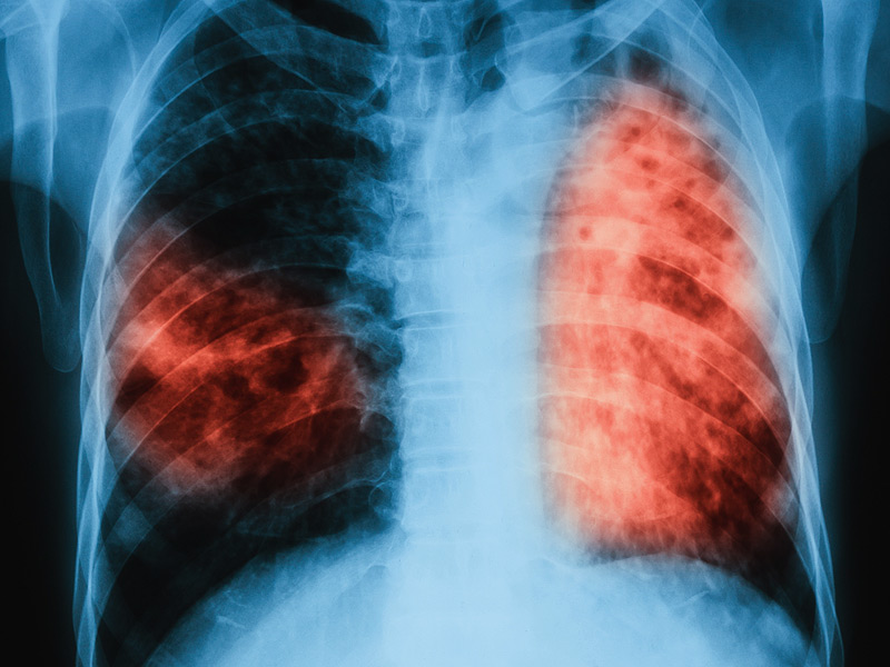
Abstract: Improved knowledge regarding the tissue penetration of antituberculosis drugs may help optimize drug management. Patients with drug-resistant pulmonary tuberculosis undergoing adjunctive surgery were enrolled. Serial serum samples were collected, and microdialysis was performed using ex vivo lung tissue to measure pyrazinamide concentrations. Among 10 patients, the median pyrazinamide dose was 24.7 mg/kg of body weight. Imaging revealed predominant lung lesions as cavitary (n = 6 patients), mass-like (n = 3 patients), or consolidative (n = 1 patient). On histopathology examination, all tissue samples had necrosis; eight had a pH of ≤5.5. Tissue samples from two patients were positive for Mycobacterium tuberculosis by culture (pH 5.5 and 7.2). All 10 patients had maximal serum pyrazinamide concentrations within the recommended range of 20 to 60 μg/ml. The median lung tissue free pyrazinamide concentration was 20.96 μg/ml. The median tissue-to-serum pyrazinamide concentration ratio was 0.77 (range, 0.54 to 0.93). There was a significant inverse correlation between tissue pyrazinamide concentrations and the amounts of necrosis (R = -0.66, P = 0.04) and acid-fast bacilli (R = -0.75, P = 0.01) identified by histopathology. We found good penetration of pyrazinamide into lung tissue among patients with pulmonary tuberculosis with a variety of radiological lesion types. Our tissue pH results revealed that most lesions had a pH conducive to pyrazinamide activity. The tissue penetration of pyrazinamide highlights its importance in both drug-susceptible and drug-resistant antituberculosis treatment regimens.
NOTE: These abstracts were retrieved from the U.S. National Library of Medicine website managed in collaboration with the U.S. National Library of Medicine


